Abdomen Ultrasound Training
Abdominal Anatomy and Imaging Essentials
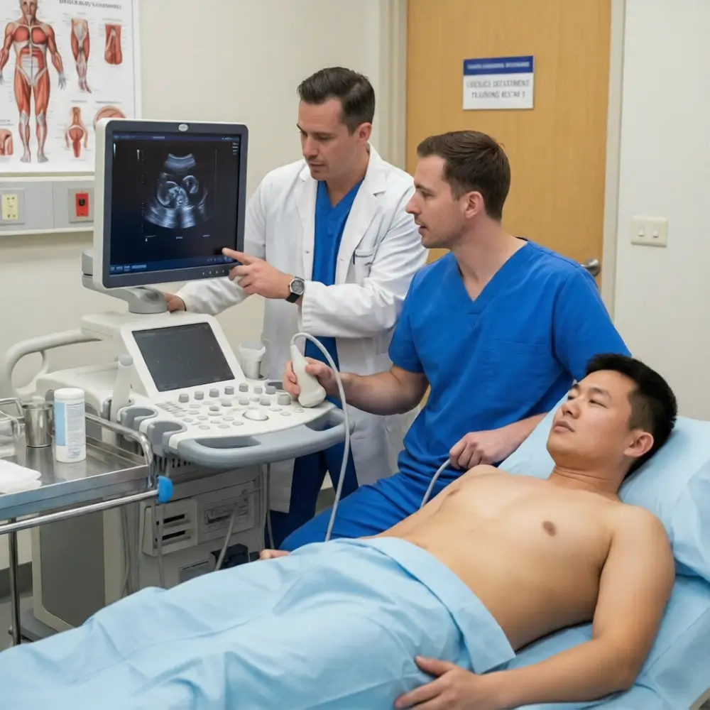
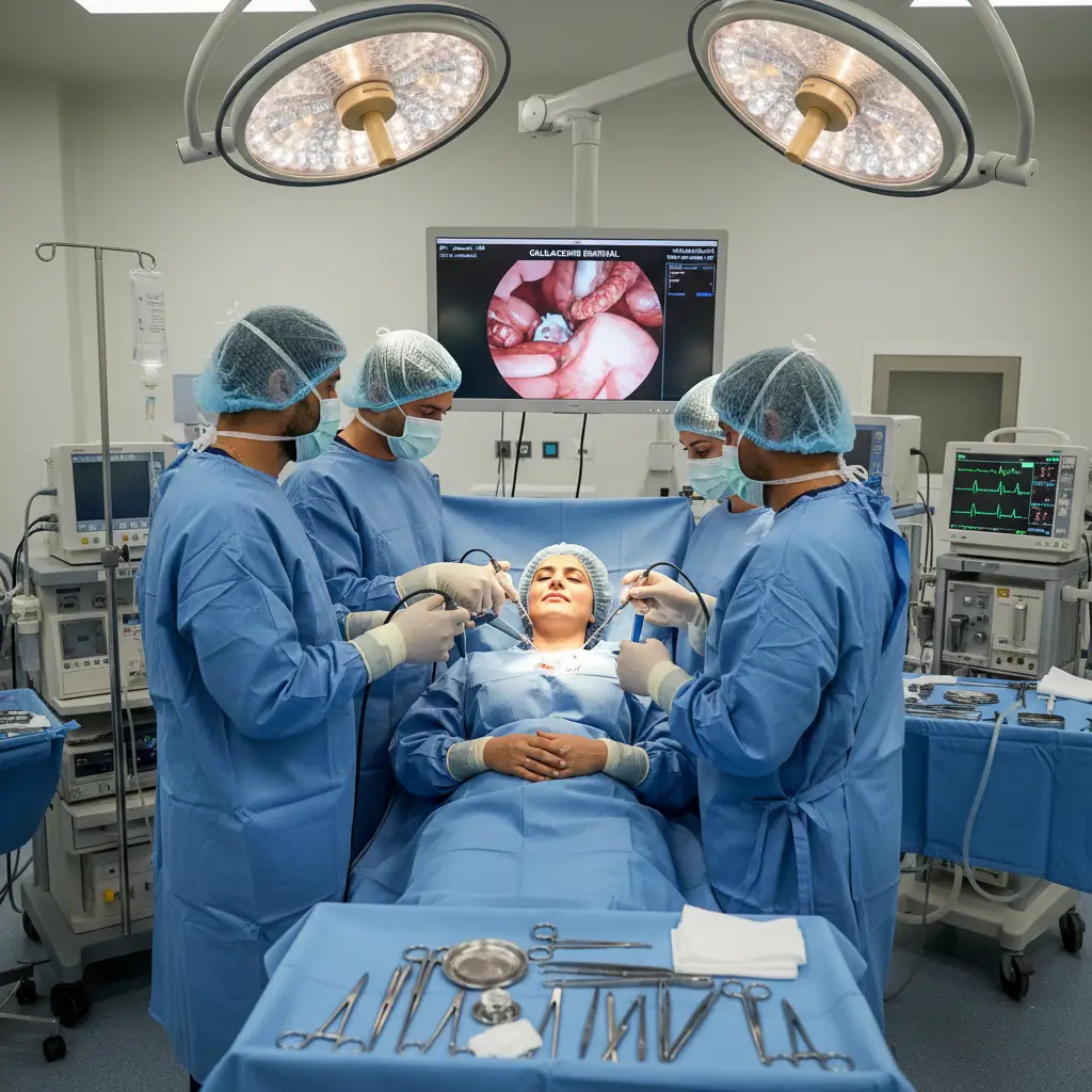
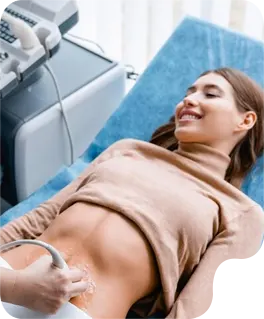
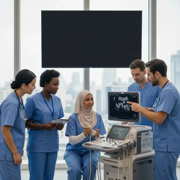
Our Abdomen Ultrasound Training is designed to help students develop a strong foundation in abdominal anatomy, scanning techniques, and image interpretation. Through structured instruction and hands-on practice, we prepare students to confidently perform abdominal ultrasound examinations in clinical settings.
As the body’s largest artery, the aorta originates from the heart and descends through the abdomen, disturbing oxygenated blood. Ultrasound is commonly used to assess the aorta for aneurysms, blockages, or other vascular conditions.
The common bile duct us a crucial tube that transport bile from the liver gallbladder to the small intestine for digestion. Ultrasound imaging helps assess the CBD for dilatation, stones, or obstruction that could impede bile flow.
The gallbladder is a small pear-shaped organ situated beneath the liver that store and concentrates bile. Ultrasound is the gold standard for visualizing the gallbladder, detecting gallstones, inflammation, or other issues affecting bile flow
The Hepatic Artery delivers oxygenated blood to the liver, providing approximately 25% of its total blood supply and a significant portion of its oxygen. Originating from the celiac trunk, it branches into the proper hepatic artery, which then divides to supply the various lobes of the liver. Distribution to hepatic artery, blood flow, such as thrombosis or stenosis, ca lead to severe liver damage due to ischemia
The hepatic veins are the primary outflow vessels of the liver, draining deoxygenated blood and filtered substances from the hepatic parenchyma into the inferior vena cava(IVC). Typically, there are three main hepatic veins-right, middle, and left-which coverage to empty into the IVC just below the diaphragm. On ultrasound, their characteristic biphasic waveform, reflecting right arterial pressure changes, is crucial for assessing liver health and detecting pathologies such as Budd-Chiari syndrome or cardiac abnormalities.
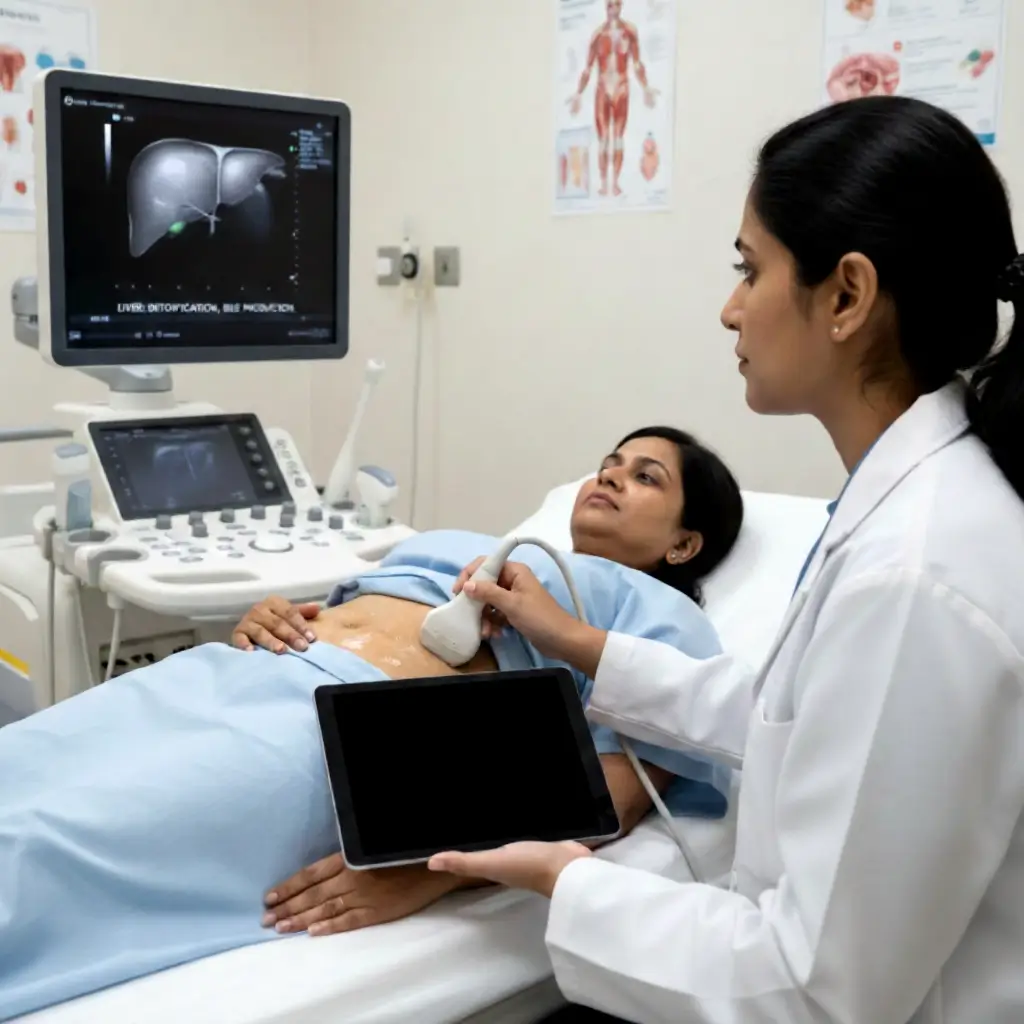
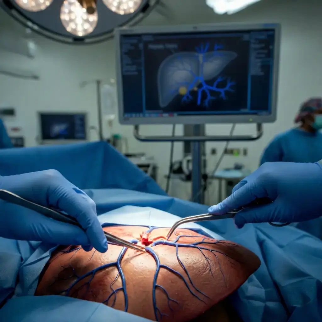
The liver is a large, complex organ in the upper right abdomen performing critical functions including detoxification and bile production. Ultrasound is a primary imaging modality for evaluating liver size, texture, and identifying masses, cysts, or signs of disease.
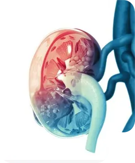
Similar to the right, the left kidney plays a vital role in maintaining fluid and electrolyte balance by filtering blood and forming urine. Ultrasound of the left kidney is invaluable for evaluating its health, detecting cysts, stones, or other abnormalities
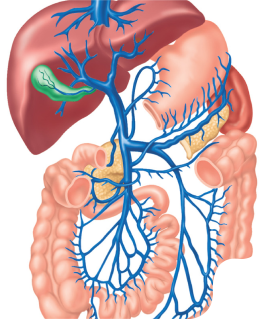
The main Portal Vein is a crucial vessel that transports nutrient-rich, deoxygenated blood from the gastrointestinal tract, pancreas, and spleen directly to the liver for processing. Formed by the confluence of the superior mesenteric and splenic veins, it provides approximately 75% of the liver’s blood supply. Understanding its normal flow and anatomy is essential for identifying various liver pathologies during ultrasound examinations.
Located in the upper left abdomen, the spleen is an organ vital to the immune system, filtering blood and storing platelets and white blood cells. Ultrasound imaging is routinely used to assess the spleen’s size, texture, and detect conditions such as enlargement or cyst.
EXCELLENTTrustindex verifies that the original source of the review is Google. I recently had the pleasure of visiting Gentle Touch Sono Lab for an ultrasound, and I cannot express how impressed I was with the entire experience. I was able to meet Ms Penn. From the moment I entered her warm, welcoming office, I felt at ease. Ms. Penn's professionalism and expertise were immediately evident, putting me at comfort during what can often be a stressful time.She took the time to explain the procedure thoroughly and answered all my questions with patience and clarity. The ultrasound itself was conducted with the utmost care, and I appreciated her attention to detail throughout the process. Her state-of-the-art equipment ensured that I received excellent images, and she made sure to share the results with me in a way that was easy to understand.What truly stood out was Ms. Penn's compassionate nature. She took the time to connect with me on a personal level, making sure I felt supported and informed every step of the way. I highly recommend Ms. Penn to anyone in need of ultrasound services. Her combination of skill, empathy, and a calming atmosphere makes her the go-to professional in this field. Thank you, Ms. Penn, for a wonderful experience!Posted onTrustindex verifies that the original source of the review is Google. Gentle Touch Sono Lab helped me enhance my skills in vascular. I was struggling with lower extremity Arterial. After a few sessions, I feel much more confident .Posted onTrustindex verifies that the original source of the review is Google. WOW. I was speechless, it was such a beautiful space filled with so many training tools. I had the pleasure of meeting Ms. Penn, she was so sweet & knowledgeable & just so passionate about the craft !! I would definitely recommend any & everyone who might be interested in sonography to go check it out.Posted onTrustindex verifies that the original source of the review is Google. I had a great experience at Gentle Touch Sono Lab. The staff was friendly, professional, and made me feel comfortable throughout the entire visit. The sonographer explained everything clearly and took their time, which I really appreciated. The lab was clean, organized, and my appointment started on time. I would definitely recommend Gentle Touch Sono Lab to anyone looking for a comfortable and professional ultrasound experience.Posted onTrustindex verifies that the original source of the review is Google. I highly recommend Gentle Touch Lab! I originally came in not knowing where to start, and Felicia made sure I received the one-on-one support I needed to excel. I’ve gained a lot of experience from both Felicia and Louis. I’ll definitely be recommending them to all my radiology classmates.
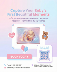
Book With Us Now
Get an Appointment
Hands-on ultrasound training for students and professionals. Bridging the gap between learning and real-world practice.
Other Links
Contact Info
- +1 888-283-6907
- info@gentletouchsonolab.com
- 41B W Merrick Rd Suite 2, Valley Stream, NY 11580, United States